
There are many ways to view a 3D animation of the heart anatomy. You can start by looking for a website that offers a 3D model of the heart that you can view from all angles. You can also modify the model to display various pathological conditions such as coronary heart disease. If you do not have a computer, you can download one for free and then animate the valves to see the effect. This way, you can learn about the heart while viewing it.
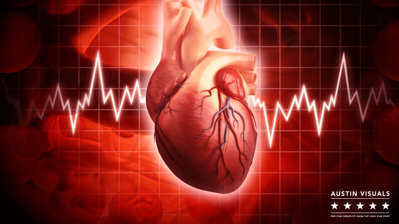
Animation of AustinVisuals’s 3D heart.
With the help of AustinVisuals, this animation shows the 3D structure of the human heart. This animation shows how electrical impulses within the heart create a normal rhythm. The two chambers of the heart pump oxygen-rich blood through valves to the body. The heart pumps oxygen-depleted blood out, while the heart returns oxygen-rich blood. This cycle is repeated 60-100 times per second.
Medically correct
An animation of the 3D anatomy of the heart will provide you with a detailed look at its structure and functions. The heart is made up of two chambers: the right heart receives deoxygenated blood from the body and pumps it to the lungs, while the left heart receives oxygenated blood and pumps it out. Blood circulates throughout the heart through veins and arteries. Brain cells die after 4-6 minutes without blood circulation. The 3D heart animation will give you an idea of how these muscles work.
To create the 3D medically accurate heart anatomy animation, thousands of static images are used to show the cardiovascular system in high-resolution. These images can be used to explain medical concepts and demonstrate surgical procedures. Medically accurate 3D animations can be used for clinical screens, courses, and more. They can break down even the most complex topics about the heart and circulatory system. Medically accurate 3D animations are available for educational purposes.
Organised
In this animated demonstration of the human heart, you can see the structures within and outside of the heart, including the membrane surrounding the atria, the prominent surface features, and the layers that form the heart wall. Each structure plays a different part in the heart’s functioning. Here, you can learn more about the heart’s structure, as well as how to recognize it and pronounce its different parts. This animation is particularly helpful for teaching children about the structure of the heart.
The pulmonary circulation is shown next, which includes the bronchial and pulmonary arteries. The pulmonary circulation pumps blood from the heart to the lungs, where it is oxygenated and returned to the heart through the pulmonary vein. The bronchial circulation moves blood from the lungs into the tissues that make up larger airways in your lungs. The first aortic arch is still intact. However, the second regresses and forms the maxillary arterial, which is located within the chest cavity.
 Organized by vascular structure
 Organized by vascular structure
An animation of the 3D anatomy of the heart can help you understand its workings. This animation will show you how blood flows through the four chambers of the heart. You will see how each chamber is constructed, including the valves. Each valve has flaps known as leaflets, which work as one-way valves. The mitral valve contains two leaflets, while the other three valves contain three. The annulus is a fibrous tissue ring that supports these flaps. It helps to maintain the correct shape of the valve. Chordae tendineae support the tricuspid and mitral valves, helping to keep the flaps in their place.
Color coding is used to represent the vascular structures of the heart. Color-coding allows for instant visual recognition and clarity. It is a useful tool for anatomy students, although students suggested that it could use less vivid colors for heart ventricles and a greater number of anatomical details could be added. In addition, the market for such software is limited, and the 3DP model only matches the anatomical details of a plastinated heart specimen.

Simple to comprehend
If you are wondering how a normal heart works, you can watch an easy to understand 3D heart anatomy animation. This animation shows you the inside workings of your heart, including the mitral and tricuspid valves as well as the aortic and tricuspid valves. These valves close and produce heart sounds. It is easy to remember and understand the 3D animation of the heart. Find out more about the heart, and how to recognize different sounds.
This 3D animated heart explains the complex structure of the heart and the interdependence of the various parts of the heart. It also helps in visualizing various diseases and procedures. You can choose from a wide variety of animations for different purposes. One of these options is a heart anatomy animation. You can choose any part of the heart that you would like to learn about to get a more comprehensive understanding of the heart.
3D Heart Anatomy Animation Services | AustinVisuals
We are a Medical Animation Company providing 3D Medical Animation, 3D Renderings, Custom Medical Graphics and Visualization services for the Healthcare profession. From Investor Pitch Videos, to Educational Content, Training, Explainer Videos for New Medical Devices, we create custom presentation Videos and still images to ensure your communication remains on point to your target audience.
Our Services
- Biomedical
- Medical Devices
- Cardiology
- Surgical
- Orthopedic
- Training / E-learning
- Character Animation
- Pharmaceutical
- Social Media / Marketing and Branding
- Explainer
- General Services
Want to know how we can help? Have questions? Have a project to discuss? Message us using the contact form below, email us [email protected] or call us (512) 591-8024 to meet with a member of our team today.